General appearance
Perforating veins corresponding to the short saphenous territory are situated in a zone between the posterolateral surface of the thigh and the bimalleolar line, which passes underneath the medial and lateral malleoli and separates the territory of perforating veins of the foot from that of the perforating veins of the ankle.
Some anatomical landmarks will help define their topography:
- the popliteal crease (in the center of the popliteal fossa);
- the cutaneous projection of the soleus muscle arch (4 fingerbreadths below the popliteal crease);
- the tip of the gastrocnemius muscles (lower limit of the fleshy part of these muscles, at the junction of the middle and lower thirds of the leg);
- the bimalleolar line.
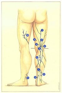

Figure 93 presents a general view of the perforating veins of the short saphenous territory.
Profunda femoris perforating veins connect a suprapopliteal trunk, constituting a continuation of the short saphenous vein on the posterior surface of the thigh, with the profunda femoris network of veins. These veins have an intramuscular course, which is longitudinal in relation to the axis of the femur.
Superficial femoral perforating veins correspond to posterior variants of Boyd’s perforating veins (see Long saphenous territory).
Perforating veins of the popliteal fossa are derived directly from the popliteal vein adjacent to the anastomosis of the common gastrocnemius vein. They perforate the fascia in the popliteal fossa.
Medial gastrocnemius perforating veins generally connect the intersaphenous vessels or the short saphenous vein itself and its collaterals to the medial gastrocnemius veins. The rich network created makes the middle segment of the short saphenous vein a crossroads for exchanges between the short saphenous. medial, and lateral gastrocnemius veins.
Gillot’s lower pole perforating vein connects a large inferior medial gastrocnemius trunk with the short saphenous vein or an intersaphenous arch. It is situated 3 to 7 cm below the tip of the gastrocnemius muscles. It can be duplicated or accompanied by a group of two to three satellite gastrocnemius perforating veins, or even by a soleus perforating vein.
Peroneal perforating veins ensure com-munication between the peroneal vein and collateral short saphenous veins, but also with collaterals derived from the long saphenous vein or infrapopliteal veins.
Medial perforating veins of the ankle are mostly situated adjacent to the malleolus, above the bimalleolar line. More posterior than Cockett’s perforating veins, they also have a smaller caliber (1 to 1.5 mm). However, they can also participate in the pathogenesis of cellulitis and leg ulcer. They connect the deep network of posterior tibial veins with a collateral short saphenous vein passing medially to tendon calcaneus or with an intersaphenous arch. They are sometimes related to the medial inframalleolar perforating veins of the foot, or more rarely with an inferior vein of the soleus muscle.
Further reading
Dortu J., Dortu JA. Les veines perforantes du membre inférieur : physiologie et physiopathologie. Phlébologie, 1994; 47: 167-75.
Gillot C. Les veines perforantes inférieures de la jambe, de la cheville et du pied. Phlébologie, 1994; 47: 76-104.
Thomson H. The surgical anatomy of the superficial and perforating veins of the lower limb. Annals of the Royal College of Surgeons, 1979; 61: 197-205.