Origins of the short saphenous vein
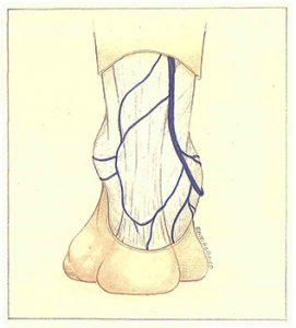
The short saphenous vein is the continuation of the lateral marginal vein of the dorsum of the foot. It then describes a curve with an anterosuperior concavity and passes underneath and then behind the lateral malleolus (Figure 89).
The lateral retromalleolar sulcus lies between the lateral malleolus and tendo calcaneus. The skin is very thin and fragile in this zone, which facilitates the development of edema. This richly innervated region is also vets sensitive. The short saphenous vein is accompanied by a sensory nerve branch, the lateral dorsal cutaneous nerve of the foot. This is derived from the sural nerve, which emerges with the short saphenous vein through the fascia cruris in the middle third of the leg.
During surgery, accidental damage to the lateral dorsal cutaneous nerve of the foot results in paresthesia of the lateral part of the heel and lateral border of the dorsum of the foot.
The origin of the short saphenous vein constitutes the saphenous portion furthest away from the heart in the standing position, and that with the highest hydrostatic pressure. Dilatation of this segment therefore constitutes an early sign of fragility of the venous wall.
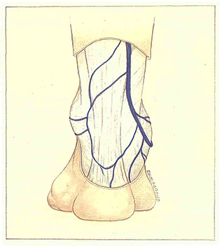
The short saphenous vein can also have a two-vessel origin (Figure 90). In this case, the main inframalleolar trunk can be accompanied by an accessory perimalleolar trunk, and the two veins anastomose just above the retromalleolar sulcus.

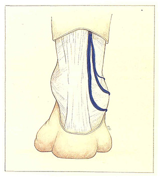
In other cases, the short saphenous vein receives a branch derived from the long saphenous vein territory, over the tendo calcaneus (Figure 91). These vessels anastomose via either a direct intersaphenous communicating vein or a periarticular venous plexus of the foot. The development of such a periarticular plexus forms a veritable perimalleolar varicose crown of anastomosis between the long and short saphenous territories.

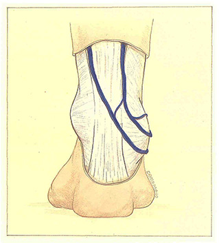
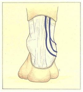
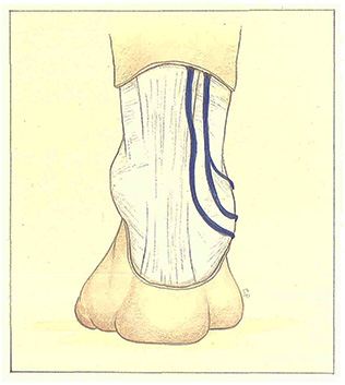
Finally, the primary trunk of the short saphenous vein can be accompanied by a parallel venous duplication (Figure 92), sometimes constituting a real accessory saphenous vein, and rejoining the main trunk at a variable level of the leg. However, this vessel may also remain independent of’ the main short saphenous vein throughout its course.
Further reading
Sobotta. Atlas d’anatomie humaine. Tome 2. Éditions Médicales Internationales, Paris, 1986.
Kahle W., Leonhardt H., Platzer W. Anatomie. Tome 1. Flammarion Médecine Sciences, Paris, 1978.