Congenital anomalies
Although rare, congenital malformations or angiodysplasias are nevertheless extremely varied.
The Klippel-Trenaunay syndrome certainly constitutes the commonest anomaly. This syndrome consists of an association of cutaneous angiomas, lengthening of the lower limb, and congenital varicose veins which therefore appear before puberty. This pure venous malformation corresponds to agenesis or hypoplasia- of the iliac vein, femoral vein or popliteal veina When it is associated with one or several arteriovenous fistulas, it constitutes the ParkesWeber syndrome.
Several elements must be investigated in any patient with angiodysplasia:
- nature of the anomaly: simple (pure venous) or mixed (arteriovenous);
- presence of agenesis or hypoplasia of a large deep venous trunk (ultrasonography) or, conversely, presence of deep dilated venous lakes or superficial megaveins;
- associated lymphatic lesions;
- presence of bone involvement or extension of the process to muscle planes;
distant malformations: inferior vena cava, renal veins, vessels of the trunk, face or upper limbs.
Many types of angiodysplasia have been described: venous aneurysms, congenital absence of valves, deep venous lakes associated with chondrodysplasia (Maffucci syndrome), venous duplications (femoral, vena cava, renal) or abnormal anastomoses. We will consider anastomosis of a deep circumflex vein into the femoral vein at the level of the saphenofemoral junction as an example of this last type of anomaly (Figure 28).
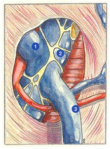
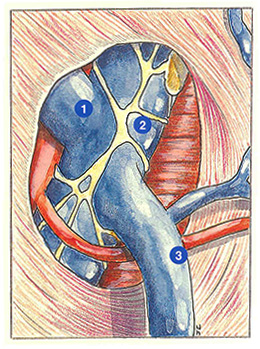
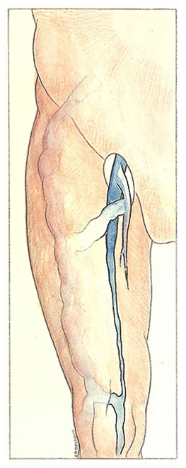
The consequences of atrophy of the femoral vein are presented in figure 29.
In this anomaly, the deep venous network of the calf is drained by a subcutaneous megavein which travels along the anterolateral surface of the leg and thigh to join the femoral vein via the arch of the long saphenous vein and the internai iliac vein via a deep circumflex or obturator vein.
This superficial megavein of the lateral surface of the thigh is frequently encountered during compression, agenesis or hypoplasia of the femoropopliteal veins.
Further reading
Browse N.L., Burnand K.G., Thomas M.L. Diseases of the veins. London, Edward Arnold, 1988: 603-25.
Klippel M., Trenaunay P. Du nxvus variqueux ostéo-hypertrophique. Arch Gen Med (Paris), 1900, 185: 641.
Shobinger R.A. Guidelines to the classification of congenital vascular malformations. Phlébologie 89, J. Libbey Eurotext, 1989: 208-20.