Clinical anatomy
Like all medical disciplines, phlebology is primarily based on clinical examination.
The data of the clinical interview are completed by those of inspection and palpation, performed with the subject standing.
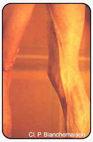
The limb examined is placed in external rotation, with the knee slightly flexed, the heel on the ground, and with muscles relaxed in order to allow palpation of the saphenous vein, accessory saphenous vein, tributaries and veins of pelvic origin.
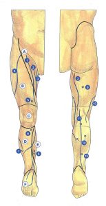
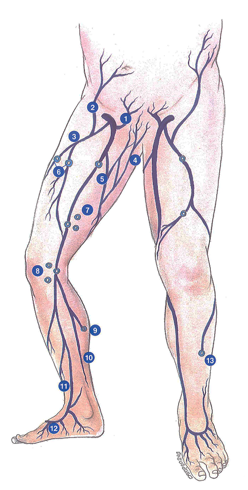
Clinically, the long saphenous territory can be divided into six anatomical regions (Figures 2 and 3).
The femoral triangle
Limited by the inguinal ligament superiorly, the sartorius muscle laterally, and the adductor longus muscle medially, the femoral triangle contains:
- the saphenofemoral junction, in the inguinal skin crease and medial to the arterial pulsation;
- the termination of the accessory saphenous veins (anterior, posterior or Cruveilhier’s vein, or parallel to the long saphenous vein in the case of deep duplication);
- the superior tributaries of the long saphenous vein – anterior branch (anterolateral vein of the thigh); – posterior branch (Giacomini’s veins which join the short saphenous vein posteriorly);
- perineal perforating veins, situated more medially and derived from pelvic veins;
- suprapubic varicose veins may also drain into the saphenofemoral junction, or even directly into the femoral vein.
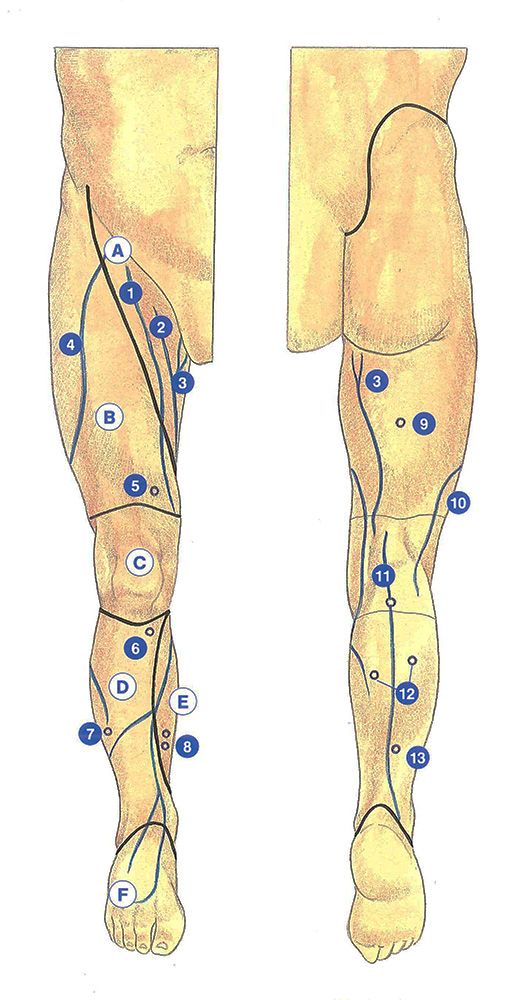
The anterior femoral region
This region, extending inferiorly as far as the knee and laterally as far as the tensor fasciae latae muscle, contains:
- the anterolateral veins of the thigh and their perforators,
- the Hunterian perforators (Dodd’s perforators),
- small perforators of the lateral surface of the thigh and venous branches derived from the popliteal fossa.
The patellar region
This region, which descends as far as the anterior tuberosity of the tibia, contains:
- the saphenous tributaries,
- certain perforators related to the anterior tributaries of the thigh, which can be detected just above tue patella.
The anterolateral leg region
Situated anterior to the tibial crest, it extends from the anterior tibial tuberosity to the tibiofibular junction.
It contains:
- the pretibial anterior saphenous tributaries,
- Boyd’s perforators at the level of the tibial tubercle,
- the peroneal perforators which are situated more laterally and which communicate with the anterolateral vein of the thigh.
The posteromedial leg region
This region, medial and posterior to the tibia, contains:
- the posterior saphenous tributaries, especially the posterior crural arch vein (described by Leonardo da Vinci) which sometimes communicates with the short saphenous vein,
- Cockett’s perforator centered around the posterior crural arch vein.
The foot region
The long saphenous vein arises from the medial marginal vein, anterior to the medial malleolus.
Further reading
Cabrol C. Anatomie. Flammarion; 1978;
Davy A. Notions d’anatomie veineuse en phlébologie quotidienne, Expansion scientifique, 1982.
Griton Ph. La maladie variqueuse, Fournier Edit., 1981.