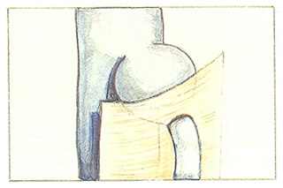
Subfascial ectasia of the saphenopopliteal junction
Anevrysmal dilatation can occur in the short saphenous vein at the saphenopopliteal junction (Figure 103).This dilatation can make a ligation flush with the popliteal vein particularly difficult.
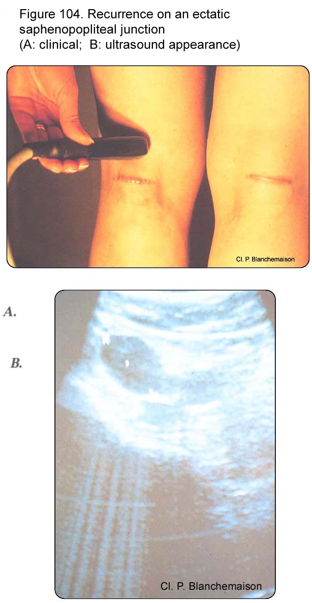
In this case, recurrence is related to an anatomical factor accentuating the hemodynamic factor. Varicose veins will then develop not on a perforating vein of the popliteal fossa independent of the short saphenous vein, but on the stump of the ectatic saphenopopliteal junction. For example, figure 104 illustrates an early recurrence, occurring 5 months after saphenopopliteal junction ligation and short saphenous vein stripping.
Further reading
Blanchemaison Ph. La physiopathologie veineuse. Phlébologie, 1995; 48: 87-8.
Davy A., Ouvry P. Recurrence of varicose veins. Phlebology, 1986; 1: 15-21.
Hobbs J. The treatment of venous disorders. MTP, Lancaster, 1977: 159-201.
Perrin M. L’insuffisance veineuse chronique des membres inférieurs. MEDSI, Paris, 1990.