Abdominopelvic veins
Pelvic veins can be the site of isolated reflux in the absence of any saphenous ,trunk involvement. They can then be responsible for real varices of the thigh or leg associated with distal reentry perforating veins.
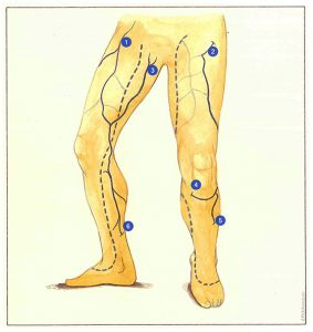
Thig situation is observed, in particular, when pelvic veins communicate with the deep venous network; for example, perineal veins (Figure 71-3) communicating with the internal pudendal or latero-uterine venous network, or, more rarely, anterior perforating veins of the groin derived from the intrapelvic venous network (Figure 71-2).
The distribution of varicose veins arising. from reflux of these pelvic veins can sometimes appear to be independent of the saphenous trunks. They drain into distal reentry perforating veins, creating veritable venovenous shunts. Several groups of perforating veins can exert this role: Boyd’s perforators (Figure 71-4), peroneal perforators (Figure 71-5) or Cockett’s perforators (Figure 71-6).
Reflux can be propagated from perineal veins to the long saphenous trunk in the upper third of the thigh, where it may supply saphenous varicose veins in the thigh and leg, while the saphenofemoral junction is also free of any valvular lesion.
In other cases, the reflux detected in the proximal part of the thigh is actually due to abdominal subcutaneous veins (Figure 71-1) ensuring countercurrent drainage of an area of abdominal skin in the absence of any deep intrapelvic communications.
Note that, from a hemodynamic point of view, the retrograde flow detected on the Doppler examination, ie reverse to that of physiological venous return, does not correspond to true reflux. It does not constitute regurgitation of blood volume from deep veins to superficial veins, but countercurrent drainage of subcutaneous venules. This phenomenon corresponds to attraction of this retrograde flow by an authentic saphenous or perineal reflux. It therefore appears important to distinguish retrograde drainage of subcutaneous tributaries from real reflux involving a venovenous shunt from deep veins to superficial veins.

Further reading
Davy A., Ouvry P., Guenneguez H.C. A propos des saphènes antérieures de cuisse. Phlébologie 1985; 38,2: 279-91.
Dortu J.A., Dortu I. Anatomie clinique du complexe saphénien à la cuisse. Phlébologie 1993; 46,1: 91-100.