Marginal veins and their branches
The role of the plantar venous pump can be more clearly understood by a precise description of the general arrangement of the veins of the foot. These veins are superimposed on four planes, from the sole upwards:
Plantar cutaneous venous plexus
This plexus is often wrongly considered to be the blood reservoir of the plantar pump. In fact, it is composed of subcutaneous and dermal venous plexuses which constitute Lejars’ venous plexus.
Medial and lateral plantar veins
These very large veins travel between the muscles of the sole of the foot and drain into the posterior tibial veins. They constitute the real blood reservoir of the plantar venous pump.
Pedal veins
These small vessels travel over the dorsal surface of the foot, between the first and second metatarsals, from the perforating vein of the first intermetatarsal space, where they communicate with the medial and lateral plantar veins, at the ankle.
Dorsal veins of the foot
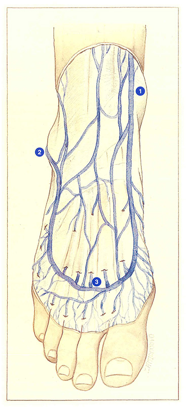
These form the dorsal venous arch, which is continuous medially with the medial marginal vein, the origin of the long saphenous vein, and laterally with the lateral marginal vein, the origin of the short saphenous vein.
The dorsal venous arch (Figure 106) is clearly visible underneath the skin. It crosses the medial segment of the five metatarsals. The convexity of this arch receives dorsal digital veins and interdigital veins, which connect, between the toes, the superficial dorsal and plantar venous plexuses (Braune’s arcade), both situated at the base of the toes. Interdigital perforating veins also anastomose in the concavity of the dorsal venous arch.
The dorsal venous arch gives rise to two or three veins of the dorsum of the foot, which sometimes merge to join the long saphenous vein or to form an accessory saphenous vein.
The dorsal venous arch continues medially as the medial marginal vein. After receiving perforating veins from the medial surface of the foot. the medial marginal vein becomes the long saphenous vein anteriorly to the medial malleolus.
Laterally, the dorsal venous arch is continuous with the lateral marginal vein, which receives perforating veins of the lateral surface of the foot. This vessel then becomes the short saphenous vein, posteriorly to the lateral malleolus.