Saphenopelvic communications
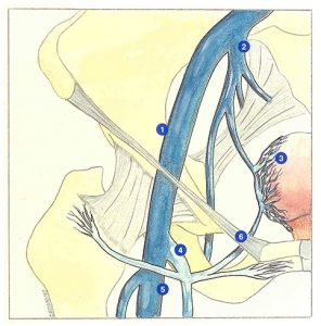
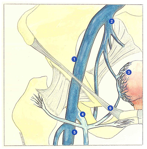
The superficial veins of the lower limbs are normally only connected to the pelvic venous system via the femoral vein which becomes the external iliac vein above the inguinal ligament (Figure 27).
The internal iliac vein or hypogastric vein is avalvular and drains all of the ipsilateral pelvic organs. In particular, if receives the uterine veins and the parietal plexus (retropubic, sacral, gluteal and obturator veins).
Ovarian veins drain into the inferior vena cava on the right and into the left renal vein on the left, while the rectal veins are drained by the inferior mesenteric vein towards the portal system.
Varicose veins of the lower limb directly supplied by intrapelvic veins communicating with the internal iliac vein are commonly observed in clinical phlebology. These varicose veins appear during a hormonal imbalance (hormonal receptors of the venous wall) or in the presence of raised intrapelvic pressure: pregnancy, pelvic venous congestion syndromes, sports requiring effort with apnea.
Three types of saphenopelvic communications can be distinguished:
- perineal perforators (Click here) which link the internal pudendal and uterine systems with the saphenous territory; qui mettent en relation les réseaux honteux interne et latéro-utérin avec le territoire saphène;
- perforators of the posterior compartment of the thigh derived from profunda femoris, ischial (caudal gluteal) or gluteal veins (Click here);
- tributaries of the arch of the long saphenous vein which communicate with obturator, uterine, or ovarian veins (Figure 27).
Selective phlebography has demonstrated this last type of communication. The presence of such variants implies considerable therapeutic consequences. For example, communication of a superficial external pudendal vein with an ovarian vein will inevitably induce a recurrence away from the saphenofemoral junction, regardless of the procedure performed (wide resection of the saphenofemoral junction, clipping, incision of the saphenofemoral junction, ligation).
Further reading
Blanchemaison Ph. Congestion et varices pelviennes. Act Med Int Angiologie, 1988, 70: 19-27.
Hobbs J.T. The pelvic congestion syndrom. Br J Hosp Med, 1990, 43: 200-6.
Kamina P., Chansigaud J.P. Anatomie fonctionnelle des veines pelviennes chez la femme. Phlébologie, 1989, 42: 363-84.